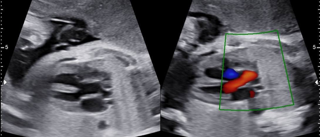
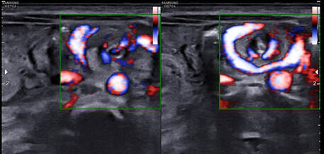
| Investigation: Ultrasound (Pregnancy) - NT Scan Diagnosis: Hypoplastic Right Heart Syndrome Gestation age 12 wk - No flow seen at tricuspid valve with echogenic appearing right veintricle. (The fetus additionally had absent nasal bone and reversed "a" wave in DV) Follow up from referring doctor - Confirmed the findings on ultrasound at 16 weeks in Chandigarh Diagnosed by: Dr Ayush Goel | 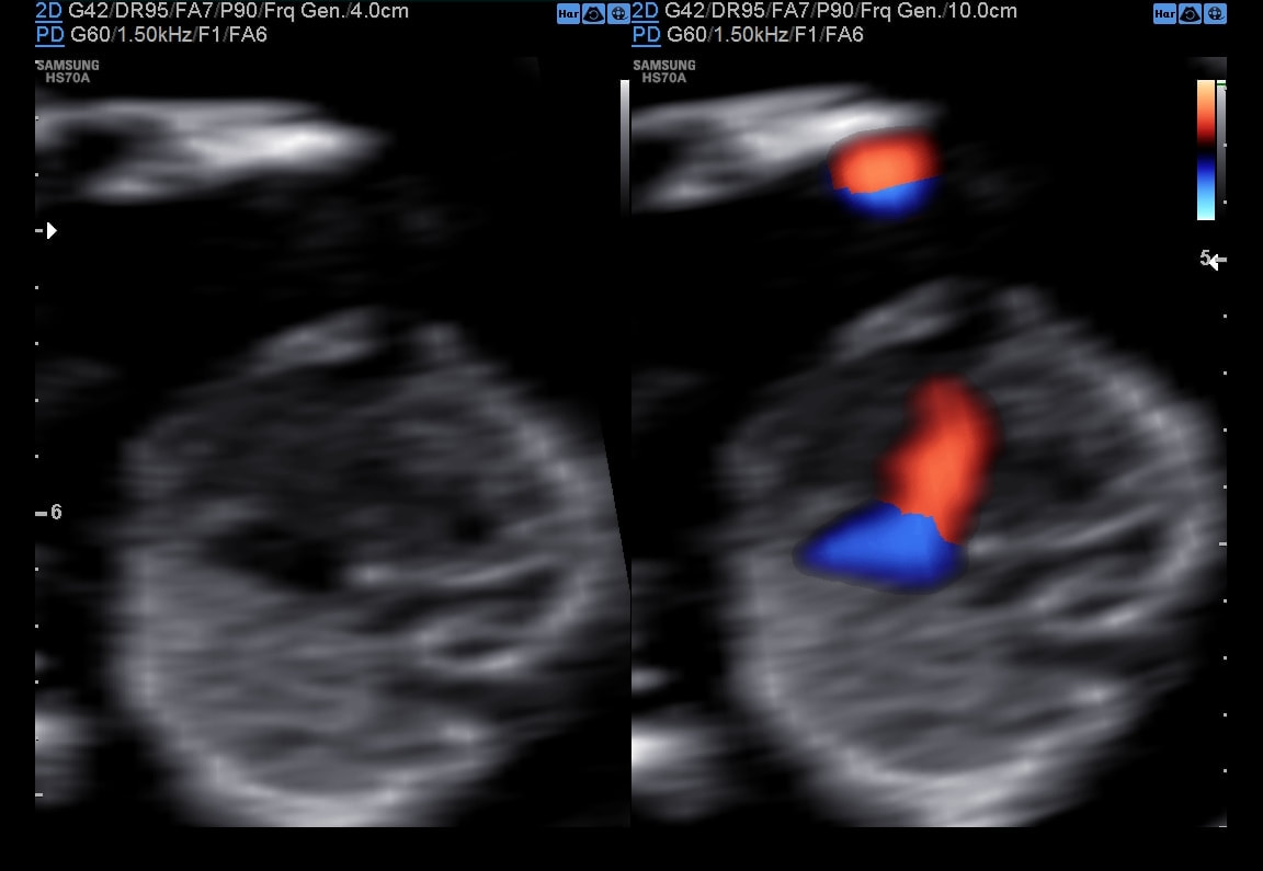 |
|
0 Comments
|
Copyrights © ShiviRadiology. All rights reserved.

Author Dr Ayush Goel
Categories
All
Subscribe To This Blog Archives Archives
January 2024
Disclaimer: The information on this BLOG is not intended or implied to be a substitute for professional medical advice, diagnosis or treatment. All content, including text, graphics, images and information, contained on or available through this blog is for medical education and enlightenment only.
While the goal of this blog is to provide complete, correct and accurate information, none of the authors/contributors can be made responsible for any incompleteness, incorrectness and inaccuracy. If you are a patient, please see your doctor for evaluation of your individual case. Under no circumstances will the authors be liable to you for any direct or indirect damages arising in connecting with the use of this blog. |
 RSS Feed
