There is always a time when a Doctor feels that his/her diagnosis significantly helped and made an important difference in patient's management.
Most of these times the patient has no idea what the Doctor went through to achieve it and how tough it was to diagnose.
Here is a log book that makes us feel proud as a Radiologist.
Note: Dates of the investigations are not disclosed to comply with Privacy Policy.
Investigation |
Diagnosis |
Images |
|
Ultrasound - FWB
Patient referred at 19-20 wks gestation for review of some anomaly not clearly defined elsewhere. |
Trisomy 13 (Patau Syndrome)
Findings: 19-20 weeks gestation with
Diagnosed by: Dr Ayush Goel |
|
|
Ultrasound - Whole abdomen
Patient referred with suspicion of hematoma in uterus in some previous scan done elsewhere |
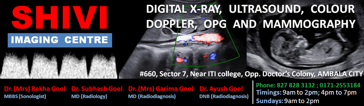
Gossypiboma
Findings: Heterogenous echogenic area distending endocervical canal with dirty shadowing and few hyperechoic specks in endometrial region - suggesting Gossypiboma. Follow up from Gynecologist confirmed Retained Sponge Gossypiboma which was removed. Diagnosed by: Dr Garima Goel |
|
|
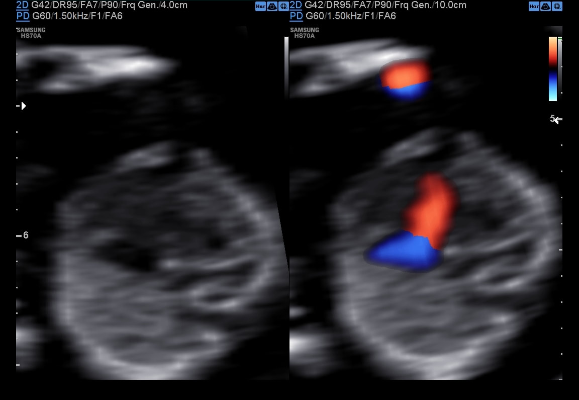
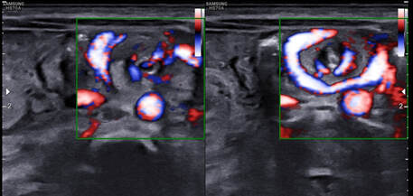
Ultrasound - Fetal Well Being
Patient came to our centre for the first time at 35 weeks gestation. |
Tricuspid atresia with VSD
Findings show: Small RV, dilated LV, Small pulmonary artery, Inlet VSD and no flow at tricuspid valve. Diagnosed by: Dr Ayush Goel |
|
|
Ultrasound (Pregnancy)- NT scan
|
Hypoplastic Right Heart Syndrome
Gestation age 12 wk - No flow seen at tricuspid valve with echogenic appearing right veintricle. (The fetus additionally had absent nasal bone and reversed "a" wave in DV) Follow up from referring doctor - Confirmed the findings on ultrasound at 16 weeks in Chandigarh Diagnosed by: Dr Ayush Goel |
|
|
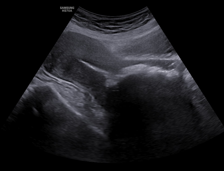
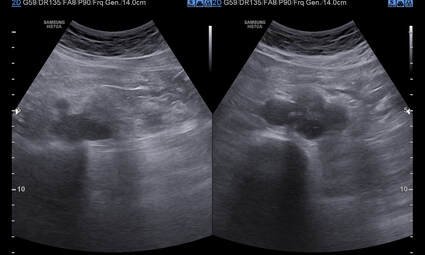
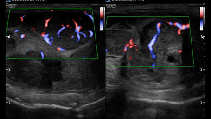
Ultrasound Whole Abdomen - female patient referred for abdominal bloating and anorexia
|
Breast Malignancy – diagnosed through metastatic ovarian lesions (Krukenberg tumor)
Bilateral ovarian masses with ascitis was seen. Liver showed multiple nodules. Patient questioned about any lump in her breast - She informed about a longstanding lump which was never evaluated. Screening of the area showed an irregular mass in breast - constellation of the imaging features are confirmative of diagnosis. Diagnosed by: Dr. Garima Goel |
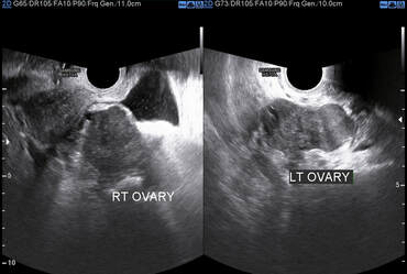
Krukenberg Tumor
Breast screened - shows typical Malignant mass
|
|
Ultrasound KUB - advised for blood in Urine
|
|
|
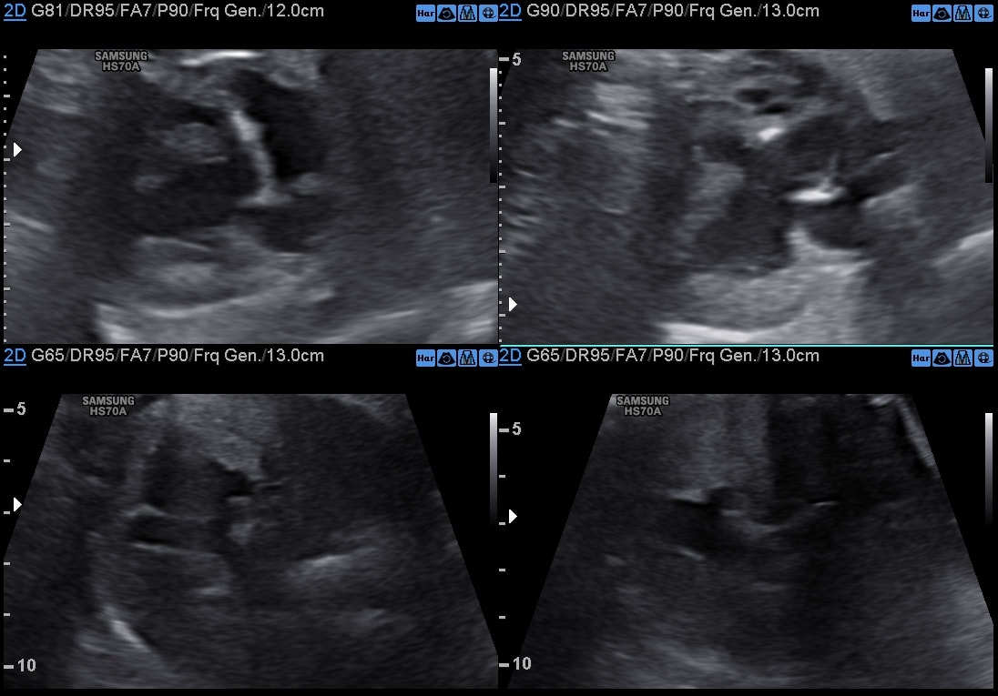
Ultrasound - Whole Abdomen -
Complains of Anorexia. |
Oesophageal malignancy
Follow up taken from the referring surgeon - endoscopy showed ulcero-proliferative growth in distal oesophagus. Diagnosed by: Dr Ayush Goel |
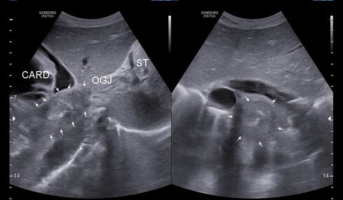
OG Junction Mass
|
|
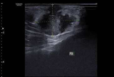
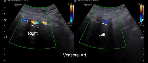
Upper Limb Arterial Doppler -
Referred for feeble pulse on left side. |
Subclavian steal syndrome
Altered phasicity was noted in left upper limb arteries. Neck was screened and reversed flow noted in Left vertebral artery. Diagnosed by: Dr Ayush Goel |
|
|
Ultrasound - Fetal Well Being
Referred for evaluation of Cerebral Ventriculomegaly |
Aberrant right subclavian artery
Apart from Ventriculomegaly, ARSA was identified, increasing the risk of Trisomy 21 by 80 times. Diagnosed by: Dr Ayush Goel |
|
|
Ultrasound Whole Abdomen -
New born with vomiting after every feed |
Midgut malrotation with Volvulus
Feedback taken from the referring doctor - the baby was immediately referred to pediatric surgeon - got operated - the child is doing well. Diagnosed by: Dr Ayush Goel |
|
|
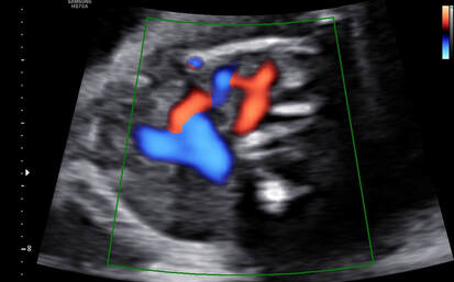
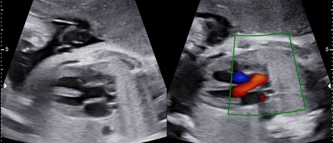
Ultrasound Fetal Well Being -
Patient came for the first time to our centre at 29 wks gestation |
Perimembranous VSD with Overriding Aorta
Diagnosed by: Dr Ayush Goel |
|
|
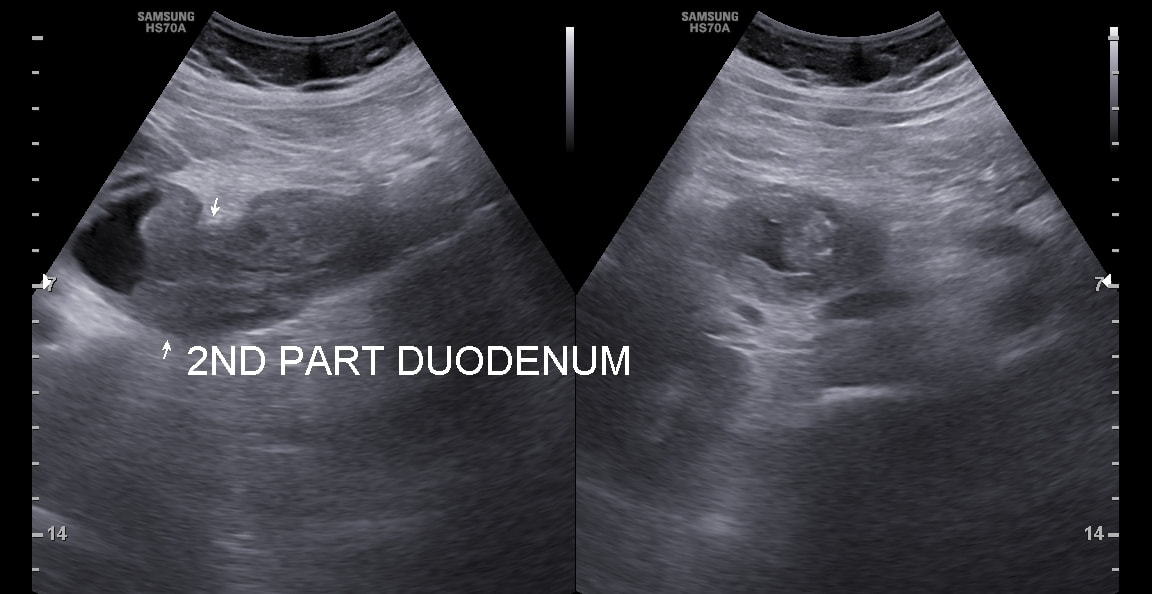
Ultrasound Whole Abdomen -
Patient complains of Vomiting |
Duodenal malignancy (early detection)
Diagnosed by: Dr Ayush Goel |
|
