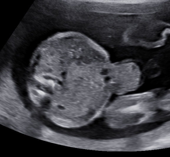
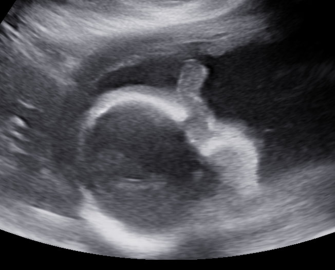
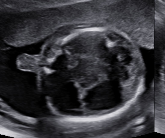
Investigation: USG FWB
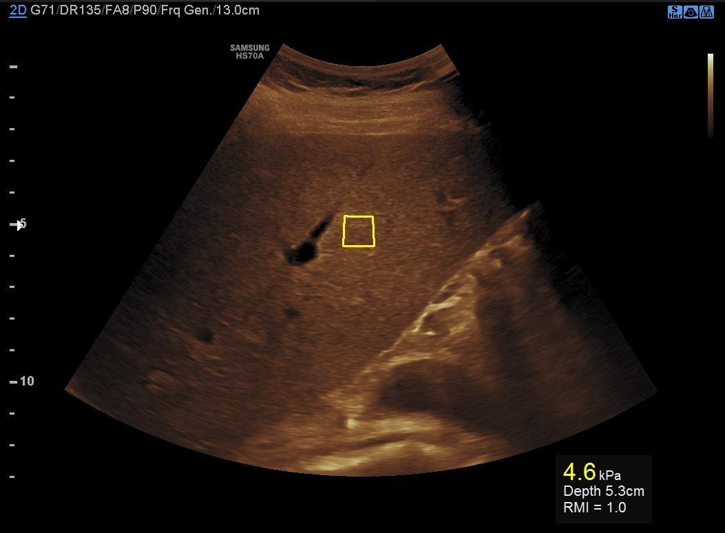
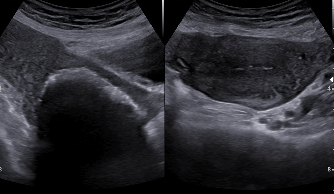
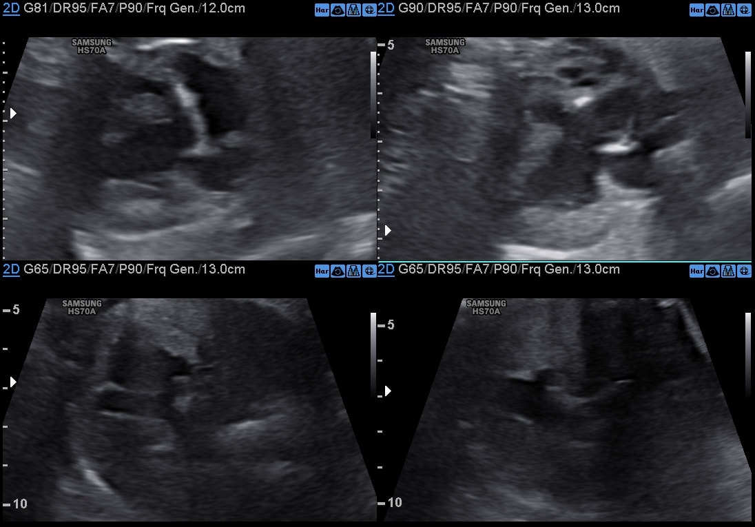
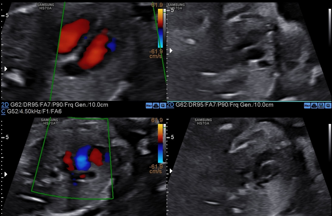
Findings: 19-20 weeks gestation with
Diagnosed by: Dr Ayush Goel
Findings: 19-20 weeks gestation with
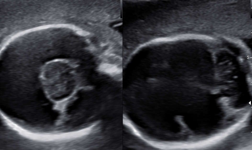
- Holoprosencephaly
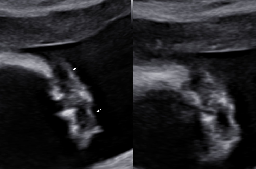
- Proboscis
- Omphalocele
- Microphthalmia & hypotelorism
- Post axial polydactyly
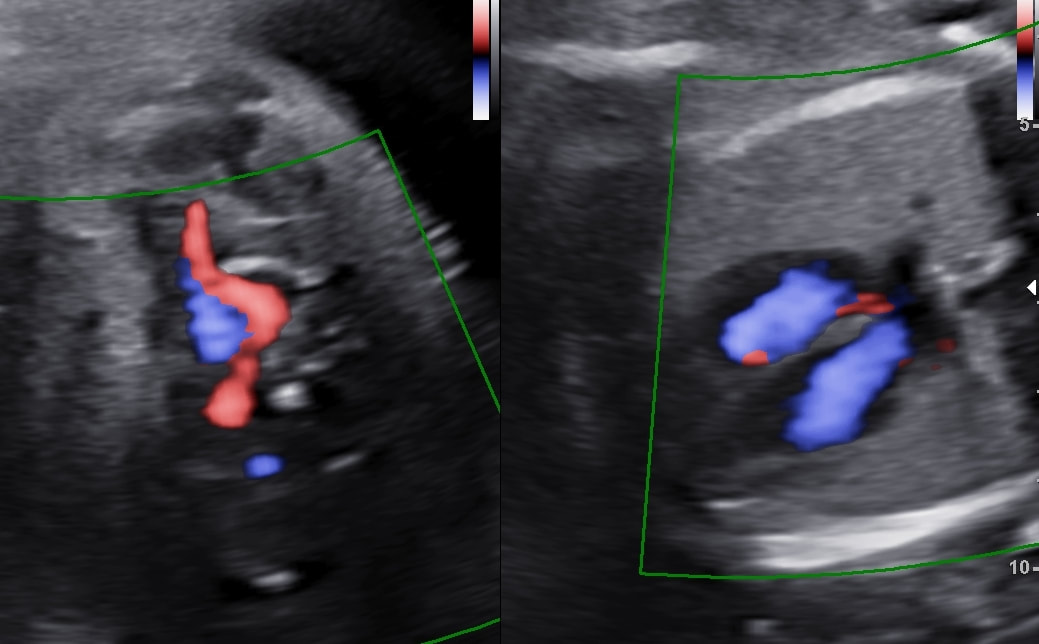
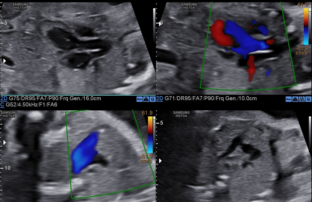
- Single umbilical artery
- Bilateral intracardial echogenic foci
Diagnosed by: Dr Ayush Goel

 RSS Feed
