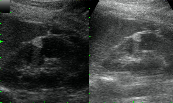
| 12 year old male presented with pain in calf and feeling of a mass in the region. Ultrasonography shows a thick walled cystic lesion with mural echogenic nodule in the medial head of gastrocnemius muscle. No abnormal vascularity was noted in the wall (not shown here). Diagnosis – Cysticercosis. Image Courtesy - DrAyushGoel |
|
0 Comments
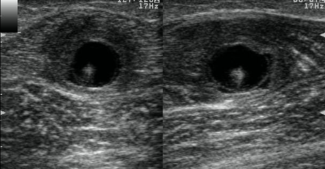
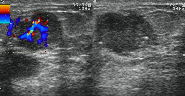
Above two images show biopsy proven malignant breast masses in two different patients.
Left image - BIRADS 5 was given with the findings (there were necrotic axillary nodes too - not shown here) Right image - BiRADS 4 was given as no axillary nodes were present but the features were suspicious) Images courtesy : Dr. Ayush Goel
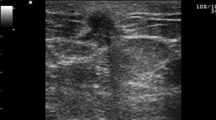
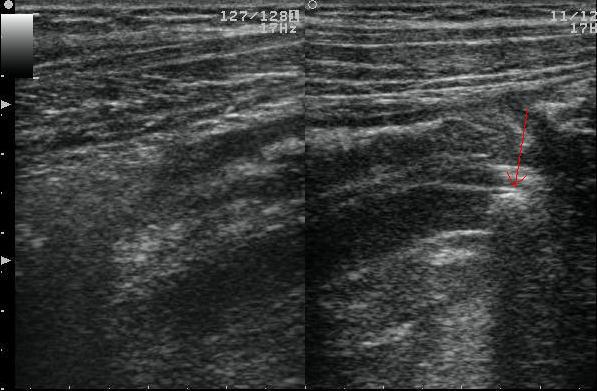
17 year old female presented with right iliac fossa pain.
USG findings - Tubular Hypoechoic, aperistaltic, non compressible, blind ending structure with an echogenic structure having posterior acoustic shadowing (red arrow) at its tip. Features are consistent with - Acute appendicitis with appendicolith. Image courtesy : Dr. Ayush Goel 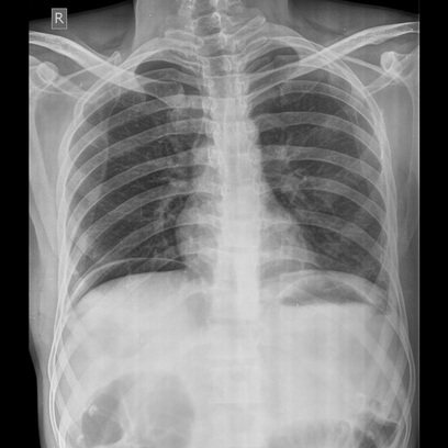 Patient presented with pain abdomen.
Erect PA chest X-ray shows air under the diaphragm (easily visible on the right, though thin lucency can be appreciated under the left dome too) Diagnosis : Pneumoperitoneum, confirmed on surgery to be due to Ileal perforation. Image courtesy : DrAyushGoel 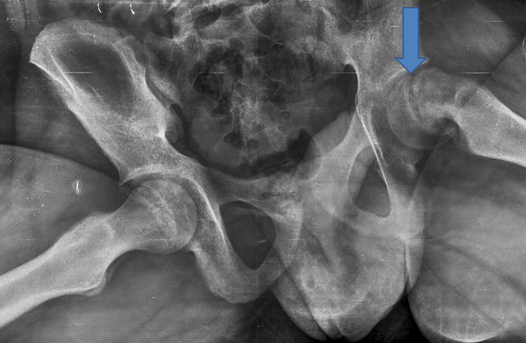 13 yr old male presents with left hip pain
Xray - Frog leg lateral view shows moderate slipped capital femoral epiphysis. Slipped capital femoral epiphysis is Type I Salter Harris fracture of epiphyseal plate. Image courtesy : DrAyushGoel |
Copyrights © ShiviRadiology. All rights reserved.

Author Dr Ayush Goel
Categories
All
Subscribe To This Blog Archives Archives
January 2024
Disclaimer: The information on this BLOG is not intended or implied to be a substitute for professional medical advice, diagnosis or treatment. All content, including text, graphics, images and information, contained on or available through this blog is for medical education and enlightenment only.
While the goal of this blog is to provide complete, correct and accurate information, none of the authors/contributors can be made responsible for any incompleteness, incorrectness and inaccuracy. If you are a patient, please see your doctor for evaluation of your individual case. Under no circumstances will the authors be liable to you for any direct or indirect damages arising in connecting with the use of this blog. |
 RSS Feed
